
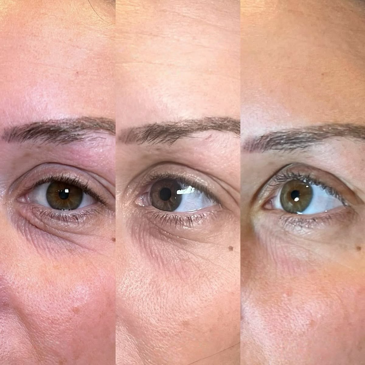
A dent under eye after filler is one of the most distressing things you can notice in the mirror a few days after your tear trough treatment. You paid for a correction. You expected a smoother, more rested appearance. Instead, there is a visible hollow — sometimes deeper-looking than what you started with — sitting right where the filler was placed.
This is one of the most common post-treatment concerns in aesthetic medicine, and the anxiety around it is completely valid. But before you call your injector in a panic, request an emergency dissolve, or conclude that the procedure failed — there is critical clinical information you need to understand first.
The dent under eye after filler that you are seeing in the first two weeks is, in the vast majority of cases, not a permanent structural problem. It is not a sign that your injector made a serious error. It is almost always a predictable, temporary phase in the healing process — one that is directly tied to how your specific tissue responds to HA filler during the under eye filler recovery time. Understanding the anatomy behind it puts you in control of your recovery instead of at the mercy of it.
What Is Actually Causing the Dent? (The Real Anatomy Behind It)
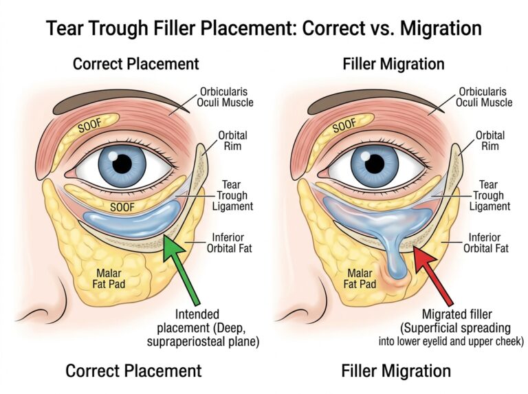
The under-eye area — specifically the tear trough — is one of the most anatomically complex injection zones on the entire face. The skin here is the thinnest on the human body, sitting directly over a network of muscles, ligaments, fat compartments, and blood vessels. When filler is placed in this region, the surrounding tissue does not respond uniformly. This uneven response is the root cause of most visible dents.
There are four distinct clinical mechanisms that can create the appearance of a dent after tear trough treatment.
1. Asymmetric Swelling Creating a False Valley
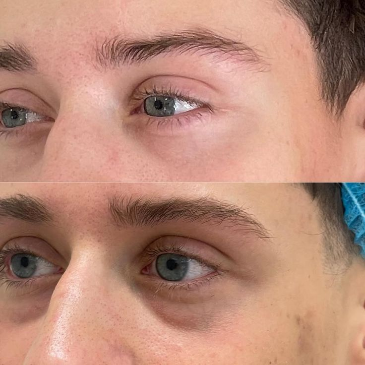
The most frequent cause of a post-filler dent is not a shortage of filler — it is actually the presence of swelling in the wrong place. When a needle or cannula enters the delicate periorbital tissue, it creates minor trauma. The body responds with localized edema (fluid accumulation). The problem is that this edema rarely distributes evenly.
If the tissue immediately adjacent to your injection site — the upper cheekbone or the orbicularis muscle just above — swells more than the injected zone itself, that swollen tissue rises above the treated area. The injected zone, which has not swollen as dramatically, now sits lower by comparison. Your brain interprets this height difference as a dent, even though the filler is perfectly placed and the volume is perfectly adequate. The dent is an optical illusion created by uneven inflammation.
This resolves completely once the swelling subsides. Patience — not intervention — is the correct response.
2. The Hydrophilic Behavior of HA Fillers
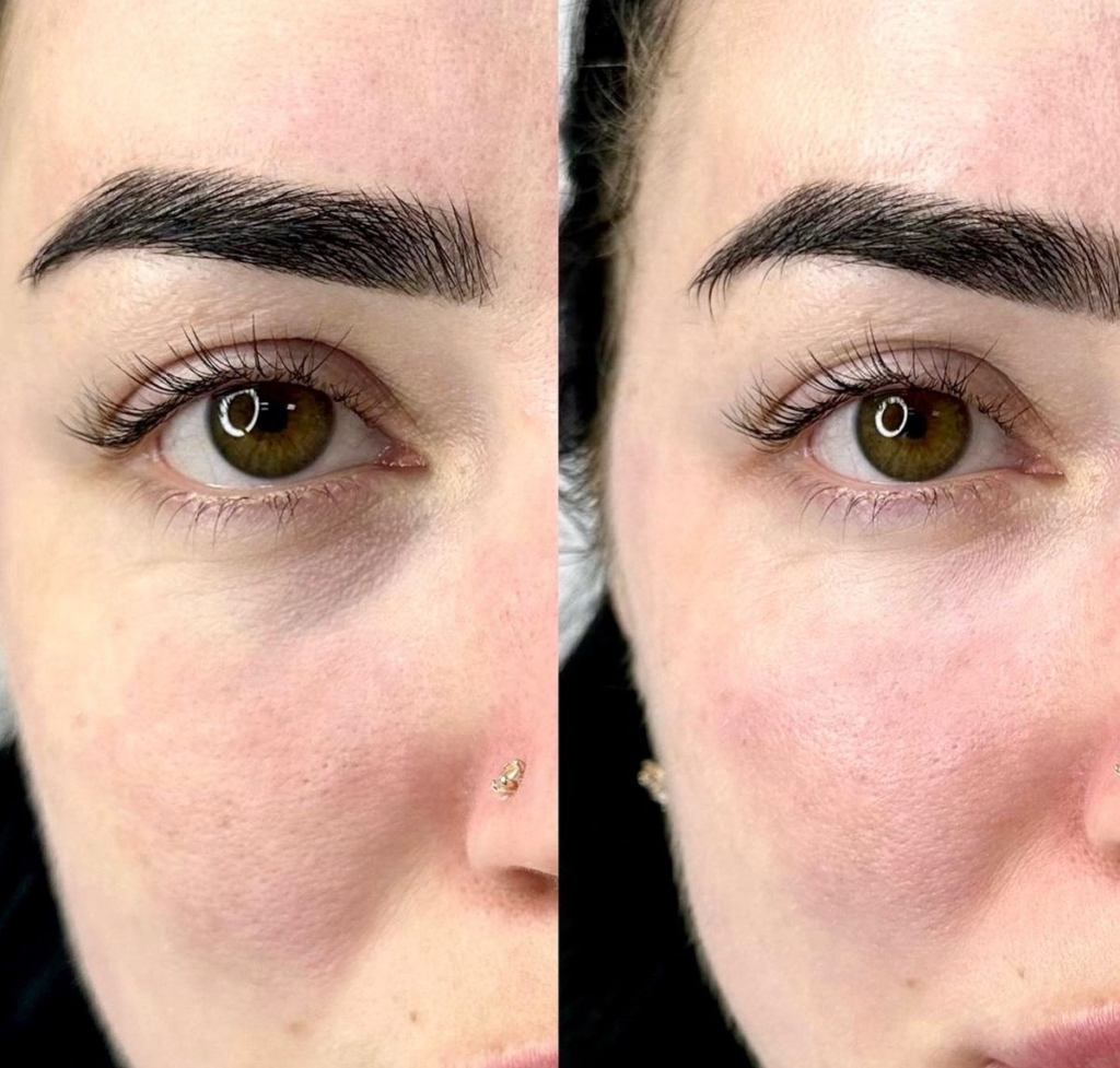
Nearly all tear trough treatments today use Hyaluronic Acid (HA) fillers such as Restylane, Juvederm Volbella, or Teosyal Redensity II. HA is a naturally hydrophilic molecule — it actively attracts and binds water molecules, sometimes holding up to a thousand times its own weight in fluid.
In the first 48 to 96 hours after injection, microscopic pockets of the filler gel absorb fluid unevenly. One small depot may hold significantly more water than the area beside it. This creates a micro-topography beneath the skin — one zone is slightly raised from fluid uptake, the neighboring zone looks comparatively sunken. The result is a shadow, a crease, or a dent-like appearance that has nothing to do with poor placement or inadequate volume.
As the HA gel reaches equilibrium with surrounding tissue hydration levels over the first two weeks, this uneven fluid distribution resolves and the surface smooths out.
3. Mid-Face Support Deficiency
This is where the anatomy gets genuinely interesting — and where a visible dent after filler can point to something structural rather than purely temporary. The tear trough does not exist in isolation. It is the junction between the lower eyelid and the upper cheek, and its appearance is heavily influenced by what is happening in the mid-face below it.
When a practitioner places filler in the cheek region to lift the malar area, that newly elevated cheek volume casts a shadow upward onto the tear trough. Depending on the precise placement — if filler is positioned slightly inferior to the cheekbone rather than along the zygoma itself — the result can be a deeper-looking hollow under the eye, not an improved one. The cheek has been raised, but the tear trough has not been bridged.
This is not a mistake unique to less experienced practitioners. The mid-face and the periorbital zone interact in ways that require three-dimensional thinking during treatment planning. Sometimes a noticeable hollow persists or worsens because the tear trough itself was not treated while the cheek was.
4. Deliberate Under-Correction
Experienced injectors working in the periorbital zone know something that first-time patients often do not: under-filling on the first session is frequently the most clinical decision a practitioner can make.
The under-eye skin has almost no tolerance for overfilling. Too much product placed too superficially results in visible lumps, the Tyndall effect (a bluish discoloration visible in natural light), or persistent puffiness that becomes difficult to manage. To avoid this, a conservative injector will intentionally place less product than the full correction would require, planning to assess the baseline result at two weeks and add more volume only if needed.
When the initial swelling clears, patients sometimes see a fraction of their original hollow still present. This is not the filler creating a dent — it is the absence of filler in that area, by design. A minor touch-up at the follow-up appointment is all that is needed.
The Recovery Timeline: When Can You Actually Evaluate Your Results?
Understanding this timeline prevents unnecessary interventions and relieves significant anxiety. Every phase of healing produces different visual results, and judging your outcome before two weeks have passed is nearly always premature.
Days 1 to 3 — Peak Trauma Response
Do not assess your results during this window. Edema and minor bruising are at their absolute maximum in the first 72 hours. The tissue is responding to the physical trauma of injection, not to the filler itself. The under-eye area can look dramatically different — puffier in some spots, deeper in others, darker from hemosiderin under the skin — and none of it reflects your final outcome.
This is also the period when HA filler is most aggressively absorbing fluid from surrounding tissue, which amplifies the uneven appearance. Many patients who contact their practitioner in distress during this window go on to have perfect, smooth results at day 14 without any intervention.
Days 4 to 13 — Integration and Flux
As the primary swelling begins to decrease, the HA gel enters what practitioners call the integration phase. The product softens, spreads slightly, and begins to interact with the surrounding dermal matrix. During this phase, the appearance of the under-eye area can fluctuate noticeably from morning to evening.
Morning puffiness often caused by lymphatic fluid pooling during sleep — can make the area look fuller but lumpier when you first wake up. By afternoon, as fluid drains, the same area may look smoother but slightly more hollow. This back-and-forth is entirely normal. The full picture of your under eye filler swelling stages only becomes clear as you move through this second week.
Day 14 and Beyond — True Baseline
The two-week mark is the universally accepted clinical standard for evaluating filler outcomes. By this point, all procedure-induced inflammation has resolved, the HA gel has fully integrated with your tissue, and the fluid dynamics have equalized. What you see now is your real result.
If the dent is gone at day 14, it was always a temporary artifact of the healing process swelling, fluid, and optical contrast doing what they do during recovery.
If the dent is still clearly visible and has been consistent for at least a week, it is a structural issue that warrants a conversation with your injector. The filler has settled. What you see is what the treatment produced.
How to Fix a Persistent Dent After Filler (Clinical Solutions)
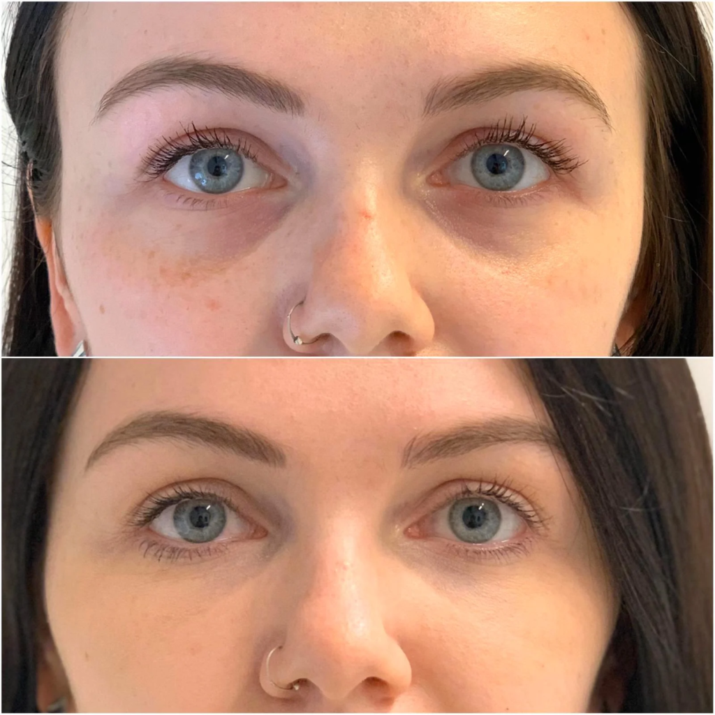
If you have passed the 14-day mark and the indentation remains, you have options. Hyaluronic Acid fillers are both malleable and reversible — this is one of the most significant clinical advantages of HA over permanent or semi-permanent alternatives. A qualified practitioner will assess your specific situation and recommend one of three approaches.
Adding Mid-Face Volume for Structural Support
If your dent stems from insufficient cheek foundation — which is extremely common, particularly in patients with significant mid-face volume loss due to aging — the solution may not be more filler under the eye. Instead, a small amount of denser, longer-lasting filler placed along the zygoma or upper cheek can lift the entire mid-face structure upward.
This lift creates a natural bridge between the cheek and the lower eyelid, smoothing the tear trough transition without adding volume directly to the most sensitive periorbital skin. It is a structurally elegant solution that experienced practitioners favor for patients with both under-eye and cheek concerns.
Precision Touch-Up for Under-Correction
If the dent is simply the result of deliberate conservative under-filling, the correction is straightforward. With the swelling completely resolved and your true baseline visible, your injector can place a micro-dose of HA product precisely into the residual hollow. The advantage of waiting until the baseline is established is that the practitioner is now working with an accurate map of your anatomy — there is no guesswork about how much to add.
This is also why responsible injectors build follow-up appointments into the treatment plan from the beginning. The first session establishes the foundation; the second session, if needed, refines it.
Dissolving with Hyaluronidase
When the problem is not too little filler but incorrectly placed filler — product that has migrated, that was deposited too superficially, or that is creating an uneven lump adjacent to the hollow — dissolution is the most effective path to correction.
Hyaluronidase is an enzyme that breaks down HA gel. According to clinical studies at the Daniel Ezra clinic, a leading oculoplastic center, hyaluronidase resolves the underlying issue in most cases, though approximately 24% of patients require more than one treatment session for complete dissolution. A small cohort — around 18% — experiences temporary hollowing or textural changes post-dissolution as the skin adjusts to the absence of the filler. This generally resolves as natural HA levels regenerate.
The critical point: hyaluronidase does not damage the skin permanently. Natural hyaluronic acid in the dermis is replenished by the body, and in most cases the treated area can be re-injected correctly after several weeks once all the dissolved filler has fully cleared.
One important caution: do not attempt to manually massage a dent or lump under the eye yourself. The periorbital skin is extraordinarily delicate. Aggressive self-massage can displace filler into unintended tissue planes and create complications that are far more difficult to address than the original issue.
When a Dent Is Not the Only Concern — Red Flags That Need Immediate Attention
Most dents and uneven areas under the eye after filler are cosmetic issues. They are distressing but not dangerous. However, certain signs warrant immediate contact with a medical professional rather than watchful waiting. Do not delay if you experience:
Blanching or Color Changes — If the skin under your eye turns white, grey, or develops a mottled purple-blue pattern, this can indicate vascular compromise — a situation where filler has inadvertently compressed or entered a blood vessel, restricting blood flow to tissue. This is a medical emergency. Contact your practitioner immediately.
Visual Disturbances — Any changes in your vision after filler, including blurring, darkening, or loss of peripheral vision in one eye, require emergency care. This is rare, but intraorbital or peri-orbital filler placement carries a theoretical risk of vascular occlusion affecting the ophthalmic artery.
Escalating Pain — Discomfort after filler injection is expected. Pain that worsens consistently over the first 24 to 48 hours rather than improving is abnormal and should be evaluated.
Signs of Infection — Increasing warmth, redness radiating from the injection site, swelling that intensifies rather than subsides, or any discharge are potential infection signs. The tear trough is anatomically close to the ocular structures; any infection in this zone needs prompt medical assessment.
Dark Circles and the Dent — A Complication Worth Understanding
There is an important distinction between a structural dent caused by the factors described above and a shadow-based dark circle that intensifies after treatment. Some patients who develop post-filler hollowing under the eye notice that their dark circles — which the treatment was meant to address — look worse, not better.
This happens because the hollow creates a shadow that reads as pigmentation. It is the same optical mechanism that causes under-eye darkness in the first place. If under eye fillers for dark circles were the primary goal of your treatment and a hollow remains, correcting the structural issue will also address the shadow-based darkness.
True pigmentary dark circles — caused by melanin deposits, visible blood vessels through thin skin, or post-inflammatory hyperpigmentation — are not affected by filler placement and require different treatments. Your practitioner can help you identify which type you have.
What Experienced Injectors Look for Before and After Treatment
The best outcomes in tear trough filler happen before the injection begins. Practitioners who achieve consistently smooth, natural results tend to share a few assessment habits:
They evaluate the patient’s malar fat pad volume, skin thickness, and tear trough ligament anatomy before placing any product, because these factors determine both the technique and the expected healing response. They discuss the realistic possibility of needing a second session — not because first-session treatment always fails, but because the conservative approach to periorbital volume replacement often requires staged correction to achieve the safest result.
They also set clear expectations about the healing timeline. A patient who understands on day one that their results will fluctuate for fourteen days is far less likely to seek unnecessary correction during the swelling phase. The anxiety that drives premature intervention is usually rooted in a lack of information about what normal recovery looks like.
The Honest Summary
A dent under the eye after filler is almost always a temporary phase in the healing process, not a permanent outcome. The two-week rule exists for a reason — it takes that long for the tissue to settle, the fluid to balance, and the HA gel to fully integrate.
If the dent persists past day 14 and is stable, it is correctable. HA filler is reversible. The tear trough is a complex area, but an experienced practitioner has a clear set of clinical tools to address whatever is causing the indentation — whether that is under-correction, mid-face support deficiency, or misplaced product.
The worst response to a post-filler dent is panic-driven intervention before the tissue has had time to heal. The best response is giving yourself the full two weeks, documenting how the area changes day by day, and then having an informed, calm conversation with your injector about what you are both seeing.
This article is intended for informational purposes only and does not substitute for personalized medical advice. Always consult a licensed, board-certified medical professional for an assessment of your specific treatment outcomes.
References
U.S. Food and Drug Administration
National Institutes of Health (NIH)

Sarah Mitchell is the founder and research editor of Under Eyes Filler, an independent patient-education resource. She is a researcher and writer, not a licensed medical professional. Her work is to read clinical studies and official FDA and ASPS guidance, then translate them into clear, honest explanations of under-eye filler results, recovery, risks, and realistic costs, so readers can prepare better questions for their own qualified provider.
[…] resolution, or how the filler has settled. If you are experiencing this, our detailed guide on what causes a dent under eye after filler explains the common causes and how it is typically […]
[…] is the honest answer: for most patients, under eye filler lasts between 9 and 18 months. That range is real, and understanding what puts you at the longer or […]